Finding Cancer in Ultrasound Images#
From pixel logistic regression to transfer learning
BreastMNIST ultrasound — metrics, trade-offs, and Grad-CAM explanations of what each model attends to
Project Overview#
Radiologists reading breast ultrasound look for irregular hypoechoic masses, angular margins, and posterior acoustic shadowing. This project asks how far a sequence of machine-learning models can get on the same task — malignant vs benign/normal — when the images are the public BreastMNIST split of the BUSI ultrasound collection.
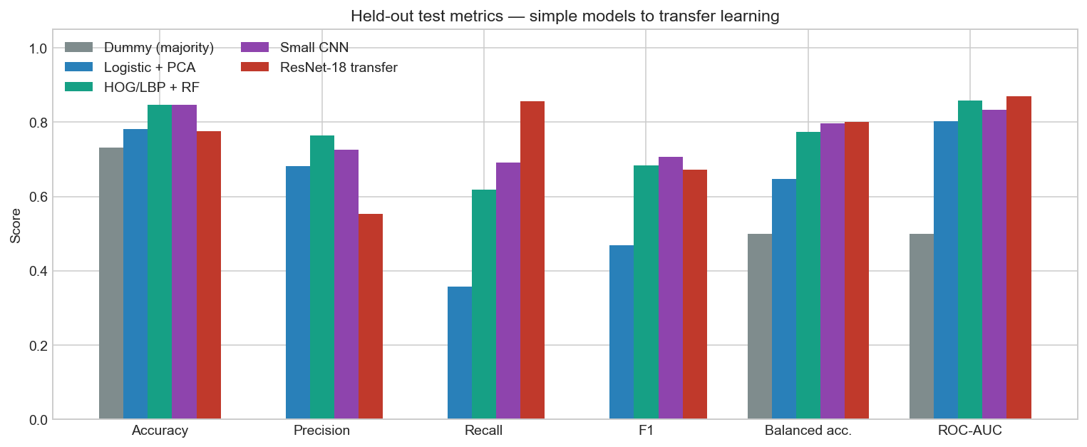
The modelling ladder is deliberate. A majority-class dummy sets the accuracy floor (never predicting cancer still scores ~73% because malignancy is the minority class). A linear model on PCA-reduced pixels is next, then classical computer-vision features (HOG and LBP) with a random forest, then a small CNN trained from scratch, and finally a frozen ImageNet ResNet-18 with a new classification head. Every model is scored on the same official test set, with decision thresholds chosen on the validation split.
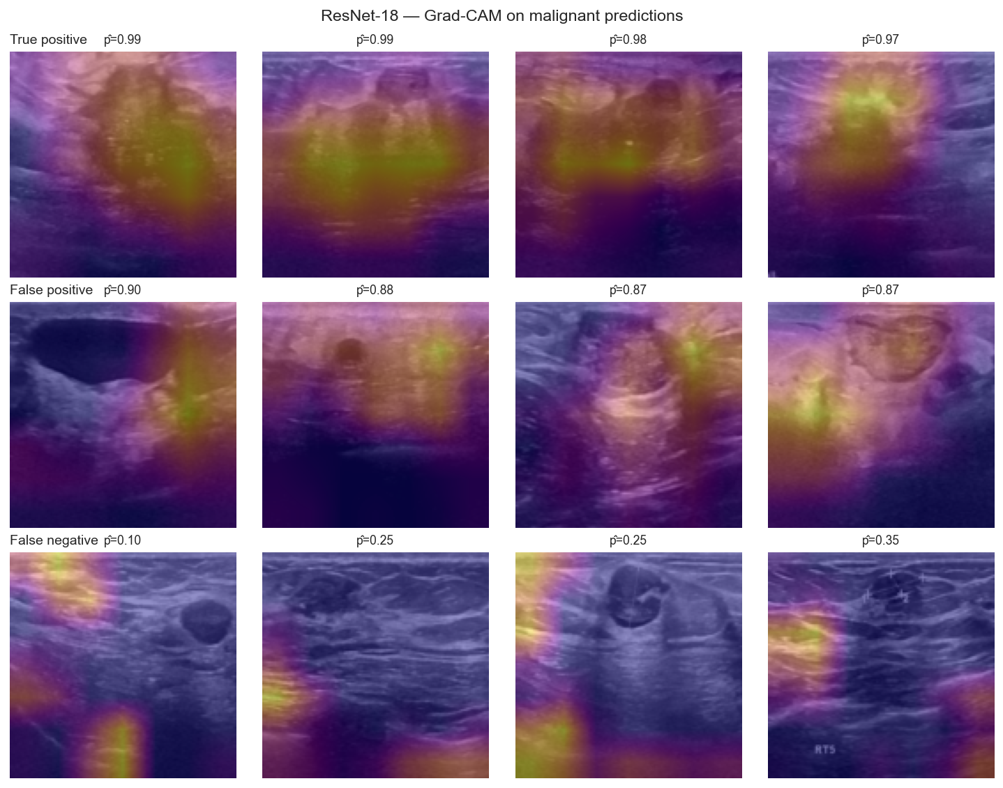
The interesting part is not a single accuracy number. Precision, recall, specificity, ROC-AUC and PR-AUC move differently as the models get more expressive — and Grad-CAM overlays show whether the conv nets are looking at the mass or at a scan artefact.
Project Structure#
Five notebooks, from the images through to a held-out comparison dashboard:
Key Questions#
If a model never predicts cancer, how high can accuracy still be — and why is that useless here?
Do hand-crafted edge and texture features beat a linear model on flattened pixels?
Does a CNN trained on 546 images outperform those classical features, or do we need transfer learning?
When Grad-CAM lights up, is it the mass, the acoustic shadow, or a measurement crosshair?
Preview Figures#
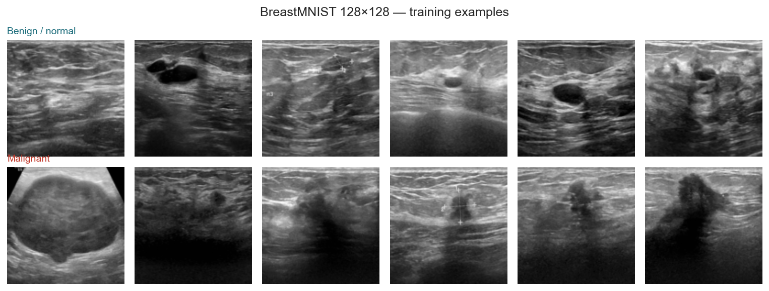
Dataset#
Source: BreastMNIST (MedMNIST v2/v3), derived from the BUSI breast-ultrasound collection
Task: binary classification, malignant = positive class (MedMNIST’s 0/1 labels are remapped in the notebooks)
Size: 780 images at 128×128 grayscale — 546 train / 78 val / 156 test
Licence: MedMNIST CC BY 4.0; cite Yang et al., Scientific Data (2023) and Al-Dhabyani et al., Data in Brief (2020)
Bundled file:
data/breastmnist_128.npz(no live download at book-build time)
Technical Stack#
Python • MedMNIST • scikit-learn • scikit-image • PyTorch • torchvision • Plotly • Grad-CAM
Ready to look at the images?